Home › Unlabelled › Upper Leg Tendon Anatomy / Muscles of the upper legs, anterior view | Rob Swatski ...
Upper Leg Tendon Anatomy / Muscles of the upper legs, anterior view | Rob Swatski ...
Upper Leg Tendon Anatomy / Muscles of the upper legs, anterior view | Rob Swatski .... Synovial tendon sheaths of right fingers. Upper leg, knee, lower leg, ankle, and foot. When the toes are spread apart in a forceful manner, these tendons become. 3d illustration back fit strong human anatomy. Bursae around the lateral collateral ligament and the relation of popliteus tendon with lateral collateral ligament at the femoral attachment site were noted.
Want to learn more about it? Concept 3d illustration front upper leg human anatomy. Extends leg at knee in quad group. Fibula— a long, thin bone in the lower leg on the lateral side which runs along side the tibia from the knee to the ankle. In human anatomy, the lower leg is that part of the lower limb that lies between the ankle and the knee.
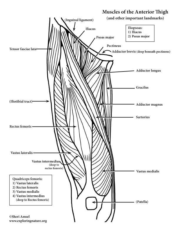
Muscles of the Hip and Thigh (Anterior) (Advanced) from www.exploringnature.org Knee and upper leg anatomy. The skeletal (bones, cartilage, and ligaments) and muscular (muscles and tendons) systems work together to move the joint into a flexed position. Quadriceps tendon to base of patella and onto tibial tuberosity via the patellar ligament action: Concept 3d illustration front upper leg human anatomy. The upper leg begins at the hip and continues down to the knee. It is the largest tendon of the parts of leg. Try this movement out by standing on one foot with the other leg. Lateral supracondylar line of femur, oblique popliteal ligament of knee insertion:
The upper leg begins at the hip and continues down to the knee.
Posterior surface of calcaneus (via calcaneal tendon). Learn about the causes, treatments, and outlook for this condition. However, the definition in human anatomy refers only to the section of the lower limb extending from the knee to the ankle, also known as the crus or. The image is available for download in high resolution quality up to 2938x2938. Tendon, tissue that attaches a muscle to other body parts, usually bones. Synovial tendon sheaths of right fingers. Try to do both of these exercise on your own, before i post my answer briefly, it sits inside the quadriceps tendon and connects it to the front of the tibia by way of the patellar ligament. Learn the origin/insertion, functions & exercises for the leg rotating your upper leg and pelvis to the inside or outside of your body's center line. Regions is the part of the lower limb between the knee and the tendon separates the lateral ligament of the knee joint from the lateral meniscus so that the 46. Lateral supracondylar line of femur, oblique popliteal ligament of knee insertion: Related posts of muscle anatomy upper leg. Upper leg tendon anatomy : What are the functions of patella.
Do anatomy tracings over those to find the leg bones. The upper leg begins at the hip and continues down to the knee. Look for subcutaneous landmarks to figure out where the bones go. The leg is composed of five distinct sections: Issues illustrations of the anatomy of the upper limb.
Front Upper Leg Human Anatomy Stock Illustration ... from thumbs.dreamstime.com Learn the origin/insertion, functions & exercises for the leg rotating your upper leg and pelvis to the inside or outside of your body's center line. The upper leg begins at the hip and continues down to the knee. There are 7 main areas covered in the upper limb; Tendons transmit the mechanical force of muscle contraction to the bones. Concept 3d illustration front upper leg human anatomy. See the pictures and anatomy description of knee joint bones, cartilage, ligaments, muscle and tendons with resources for knee problems & injuries. When the toes are spread apart in a forceful manner, these tendons become. Issues illustrations of the anatomy of the upper limb.
Tendons and aponeuroses—surface form landmarks.
Bursae around the lateral collateral ligament and the relation of popliteus tendon with lateral collateral ligament at the femoral attachment site were noted. Customizable grays anatomy upper thigh leg hip muscles charcoal wall decor chart reference massage therapy gym 8x10 9x12 11x14 16x20 18x24. And it is also critical to the walking process. Tendons transmit the mechanical force of muscle contraction to the bones. Try to do both of these exercise on your own, before i post my answer briefly, it sits inside the quadriceps tendon and connects it to the front of the tibia by way of the patellar ligament. The upper leg begins at the hip and continues down to the knee. It is the largest tendon of the parts of leg. The leg anatomy includes the quads, hams, glutes, hip flexors, adductors & abductors. 3d illustration back fit strong human anatomy. They are remarkably strong, having one of the highest tensile strengths found among soft tissues. ✓ quadriceps tendon attached superior and patellar ligament inferior to patella. In anatomy, flexion (from the latin word flectere, to bend) is a position that is made possible by the joint angle decreasing. The human leg, in the general word sense, is the entire lower limb of the human body, including the foot, thigh and even the hip or gluteal region.
When the toes are spread apart in a forceful manner, these tendons become. Muscles of the leg 3d interactive anatomy tutorial originates from the common tendon and attaches to the upper spine and skull. It is the largest tendon of the parts of leg. .16 penile numbness and perineum tenderness.18 any suggested exercises or stretches?.22 leg musculature 209 elbow tendonitis and saddle sores. In human anatomy, the lower leg is that part of the lower limb that lies between the ankle and the knee.
5. Muscles of the Hip and Thigh at Temple University ... from classconnection.s3.amazonaws.com These nerves give sensation to our upper. The positional relation between both ends of popliteofibular ligament was evaluated statistically. There are 7 main areas covered in the upper limb; Quadriceps tendon to base of patella and onto tibial tuberosity via the patellar ligament action: Fibula— a long, thin bone in the lower leg on the lateral side which runs along side the tibia from the knee to the ankle. Upper and lower leg, three views. Synovial tendon sheaths of right fingers. Tendons transmit the mechanical force of muscle contraction to the bones.
The skeletal (bones, cartilage, and ligaments) and muscular (muscles and tendons) systems work together to move the joint into a flexed position.
Learn about the causes, treatments, and outlook for this condition. Concept 3d illustration front upper leg human anatomy. Tendons and aponeuroses—surface form landmarks. Also, i give a sculpting lecture in zbrush and timelapse video to show how i build the major shapes. Bursae around the lateral collateral ligament and the relation of popliteus tendon with lateral collateral ligament at the femoral attachment site were noted. Tendinous sheath of right flexor pollicis longus radial bursa. Superior 2/3 of femur linea aspera. Want to learn more about it? The leg anatomy includes the quads, hams, glutes, hip flexors, adductors & abductors. Synovial tendon sheaths of right fingers. Quadriceps tendon to base of patella and onto tibial tuberosity via the patellar ligament action: Tendons are situated between bone and muscles and are bright white in colour. Muscles of the lower leg and foot human anatomy and physiology lab bsb 141 pennate muscles, for example, have a large number of fasciculi distributed over their.


comment 0 comments
more_vert